
Leukemia: Symptoms, Types, Causes, and Treatment Options (Ethiopian Patient Guide 2026)
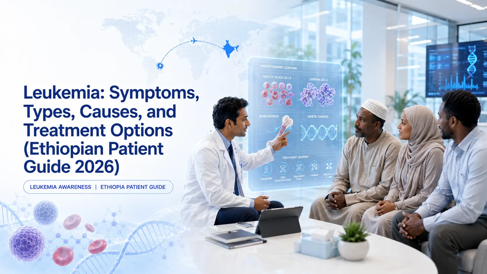
Leukemia is a cancer of the blood-forming tissues in the bone marrow, classified into four main types (ALL, AML, CLL, CML) based on speed of progression and cell type affected. Common symptoms include persistent fatigue, frequent infections, easy bruising, bone pain, and unexplained weight loss. Causes range from genetic factors to environmental exposures. Treatment options include chemotherapy, targeted therapy, immunotherapy, and bone marrow transplant.
Why This Guide Matters for Ethiopian Patients
Leukemia is one of the most common cancers diagnosed in Ethiopia, particularly at Tikur Anbessa Specialized Hospital and Bethel Teaching Hospital. Many Ethiopian patients first encounter information about leukemia after a worrying complete blood count (CBC) result, a doctor's mention, or a family member's diagnosis.
Understanding what leukemia is, how it behaves, and what causes it removes much of the fear that comes with a diagnosis. This guide is built to be a complete first reference for Ethiopian patients and families researching leukemia, written in plain language without unnecessary medical jargon.
This guide covers what leukemia is, the four types, symptoms to watch for, causes and risk factors, how it is diagnosed, and treatment options including the role of advanced treatment available in India for Ethiopian patients.
What is leukemia?
Leukemia is a cancer of the blood-forming tissues in the bone marrow, characterized by the uncontrolled production of abnormal white blood cells that crowd out the healthy blood cells the body needs to function.
Blood does three critical jobs in the body. Red blood cells carry oxygen from the lungs to organs throughout the body. White blood cells fight infections from bacteria, viruses, and fungi. Platelets help blood clot when there is bleeding from cuts or injuries. All three are produced in the bone marrow, the soft tissue inside larger bones such as the hip, sternum, and thigh bones.
In a healthy body, bone marrow produces these cells in a tightly controlled process. Stem cells in the bone marrow gradually mature into the three different blood cell types, then enter the bloodstream to do their work. Old cells die off and new cells replace them at a steady, controlled pace.
Leukemia disrupts this process. Abnormal white blood cells form due to DNA damage in the bone marrow stem cells. These abnormal cells multiply uncontrollably. They take up space in the bone marrow that should be used to produce healthy red blood cells, normal white blood cells, and platelets. Over time, the bone marrow becomes so crowded with abnormal cells that it cannot produce enough healthy cells to keep the body functioning.
This is why leukemia causes its characteristic symptoms. Low red cell counts cause anemia and persistent fatigue. Low normal white cell counts cause frequent infections. Low platelet counts cause easy bruising, bleeding gums, and nosebleeds.
The Amharic term for leukemia is የደም ካንሰር (yedem kānsar), meaning blood cancer. This is the most common name used in Ethiopia.
Leukemia is different from solid tumor cancers such as breast cancer or lung cancer. Solid tumors form a single mass that can be removed surgically. Leukemia does not form a mass; it spreads through the blood from the start, which is why surgical removal is not possible and treatment is built around medications that reach the entire bone marrow and bloodstream.
How does leukemia affect the body?
Leukemia affects the body by replacing healthy blood cells with abnormal white blood cells, causing fatigue from anemia, infection susceptibility from immune system failure, bleeding from low platelets, and organ infiltration as leukemia spreads.
Effect on red blood cells
Healthy red blood cells carry oxygen from the lungs to organs throughout the body. When bone marrow space is taken over by leukemia cells, red blood cell production drops. The result is anemia, which causes persistent fatigue, shortness of breath with mild exertion, pale skin, and weakness. Severe anemia in advanced leukemia often requires blood transfusions during treatment to maintain organ function.
Effect on normal white blood cells
Healthy white blood cells fight bacterial, viral, and fungal infections. Leukemia produces abnormal white cells that look like white cells under a microscope but cannot fight infection. The body's CBC may show very high white blood cell counts, yet the patient has essentially no functional immunity. The result is frequent or severe infections that take a long time to clear. This is often the symptom that prompts a leukemia diagnosis, particularly when an Ethiopian patient has received repeated antibiotic treatment for "recurrent infections" without identifying the underlying cause.
Effect on platelets
Healthy platelets help blood clot to stop bleeding from minor injuries. Low platelet counts in leukemia cause easy bruising, including bruises that appear without any injury, nosebleeds, bleeding gums when brushing teeth, and prolonged bleeding from minor cuts. Petechiae, which are tiny red dots under the skin that do not blanch when pressed, are a particularly important sign of low platelets. Severe platelet drops in advanced leukemia can cause spontaneous bleeding, sometimes requiring platelet transfusions during treatment.
Effect on organs
As leukemia advances, abnormal cells can spread beyond the bone marrow. The spleen and liver often enlarge as leukemia cells accumulate, sometimes causing a feeling of fullness or discomfort in the upper abdomen. The central nervous system can be involved, particularly in acute lymphoblastic leukemia (ALL), causing headaches and other neurological symptoms. The testes can be involved in male ALL patients. Lymph nodes can enlarge, particularly in chronic lymphocytic leukemia (CLL). Skin and gum involvement can occur in acute myeloid leukemia (AML), sometimes appearing as raised skin lesions or gum swelling.
What are the four types of leukemia?
Leukemia is classified into four main types based on two factors: how quickly it progresses (acute or chronic) and which white blood cell type is affected (lymphoid or myeloid). The four types are Acute Lymphoblastic Leukemia (ALL), Acute Myeloid Leukemia (AML), Chronic Lymphocytic Leukemia (CLL), and Chronic Myeloid Leukemia (CML).
Acute Lymphoblastic Leukemia (ALL)
ALL is the most common cancer in children worldwide, with a peak incidence between ages 2 and 5 and a smaller peak in older adults. It affects lymphoid white blood cells and progresses rapidly without treatment.
Common presentation in Ethiopian children includes persistent low-grade fever, bone pain that may cause a child to limp or refuse to play, fatigue, and easy bruising. The most treatable childhood cancer when full protocol is accessible, ALL has cure rates of 75 to 90% in children depending on access to comprehensive treatment.
ALL has two main subtypes based on cell origin. B-cell ALL is the most common subtype. T-cell ALL is less common and tends to occur in adolescents and young adults.
Acute Myeloid Leukemia (AML)
AML is the most common acute leukemia in adults and increases in frequency with age. Most diagnoses occur after age 50, though AML can occur at any age. It affects myeloid white blood cells and progresses rapidly without treatment, sometimes within weeks.
Common presentation includes severe fatigue, frequent infections, easy bleeding, and small red spots under the skin. Cure rates with full treatment range from 35 to 65% depending on age, subtype, and access to bone marrow transplant.
AML has multiple subtypes (M0 through M7 in the older FAB classification, with newer classifications based on genetic features). Acute Promyelocytic Leukemia (APL, M3) is a distinct AML subtype with very high cure rates using targeted therapy with all-trans retinoic acid (ATRA) and arsenic trioxide.
Chronic Lymphocytic Leukemia (CLL)
CLL is the most common chronic leukemia in older adults, with median diagnosis age around 70 years. It affects mature lymphoid cells (typically B-cells) and progresses slowly, often over many years.
CLL is often discovered incidentally on routine blood tests in patients with no symptoms. Many CLL patients do not require immediate treatment and are managed with a "watch and wait" approach until symptoms develop or blood counts deteriorate significantly.
Symptoms develop gradually and include enlarged lymph nodes that are typically painless, fatigue, frequent infections, and enlarged spleen. CLL is less common in Ethiopia historically but is increasing as life expectancy rises.
Chronic Myeloid Leukemia (CML)
CML affects myeloid cells in chronic phase. Average diagnosis age is 50 to 60 years. The defining feature is the Philadelphia chromosome, a specific genetic abnormality formed when parts of chromosomes 9 and 22 swap places, creating the BCR-ABL gene fusion that drives CML. Over 95% of CML cases have this genetic feature.
CML progresses slowly over years if untreated. Modern treatment with tyrosine kinase inhibitors (TKIs) such as Imatinib (Gleevec), Dasatinib, and Nilotinib has transformed CML from a previously fatal disease into a manageable chronic condition. Most patients on TKI therapy today live with normal life expectancy.
CML has three phases. Chronic phase, where most patients are diagnosed, is the slow-progressing form. Accelerated phase is a transition stage where the disease becomes harder to control. Blast crisis resembles acute leukemia and requires aggressive treatment. The goal of TKI therapy is to keep patients in stable chronic phase indefinitely.
The cost of TKI therapy in India is dramatically lower than Western alternatives, making lifetime treatment realistic for Ethiopian patients who can access Indian medication sources.
What is acute vs. chronic leukemia?
Acute leukemia progresses rapidly with immature abnormal cells requiring immediate treatment, while chronic leukemia progresses slowly with more mature cells often allowing extended treatment-free periods or long-term management with daily medication.
Acute leukemia characteristics
Acute leukemia progresses rapidly with symptoms developing over weeks to months. The abnormal cells are immature, called blasts, and cannot perform any normal function. Without treatment, acute leukemia can be fatal within weeks to months.
Acute leukemia requires immediate treatment, often within days of diagnosis. Treatment is intensive, involving multiple phases of chemotherapy and sometimes bone marrow transplant. The standard treatment timeline runs 2 to 3 years total when consolidation and maintenance phases are included. Examples are ALL and AML.
Chronic leukemia characteristics
Chronic leukemia progresses slowly with symptoms developing gradually over months to years. The abnormal cells are more mature and retain partial function. Some patients with chronic leukemia can live for years before treatment becomes necessary.
Chronic leukemia treatment can be daily oral medication (the standard approach for CML with TKIs) or chemotherapy combined with targeted therapy (for advanced CLL). Examples are CLL and CML.
Why this matters for treatment urgency
Patients diagnosed with acute leukemia in Ethiopia need urgent referral and treatment decision within days to weeks. The condition does not allow time for extended deliberation. Patients diagnosed with chronic leukemia have more time for second opinions, treatment planning, and family decisions. DocTrePat coordinates expedited 7 to 10 day travel for acute leukemia cases requiring urgent treatment in India when this timeline is medically necessary.
What are the symptoms of leukemia?
Leukemia symptoms include persistent fatigue, frequent infections, easy bruising or bleeding, bone or joint pain, swollen lymph nodes, fever, unexplained weight loss, pale skin, night sweats, and tiny red spots under the skin (petechiae). These symptoms are often misdiagnosed as anemia, typhoid, or other common infections in Ethiopia.
General symptoms across all leukemia types
Common leukemia symptoms include persistent fatigue that does not improve with rest, frequent or severe infections (chest infections, sinus infections, skin infections that take a long time to heal), easy bruising including bruises that appear without injury, bleeding gums or nosebleeds, prolonged bleeding from minor cuts, bone or joint pain (particularly in children with ALL), swollen lymph nodes in the neck, armpits, or groin, persistent low-grade fever, unexplained weight loss, pale skin (a sign of anemia), night sweats, petechiae (tiny red dots under the skin that do not blanch when pressed), shortness of breath with mild exertion, and reduced appetite.
Symptoms specific to acute leukemia (ALL and AML)
Acute leukemia symptoms develop rapidly, usually over weeks. Severe fatigue worsens daily. Recurrent infections do not clear with standard antibiotics. New bruises appear without cause. Children may refuse to walk or play due to bone pain. Adults may report a new feeling of being "knocked off their feet" by minor illnesses.
Symptoms specific to chronic leukemia (CLL and CML)
Chronic leukemia symptoms develop slowly over months to years. Some patients have no symptoms initially, with CLL often detected on routine blood tests. CML may present with a feeling of fullness or discomfort on the left side of the abdomen due to an enlarged spleen. CLL may present with painless enlarged lymph nodes that the patient or family notices but doesn't initially associate with serious illness. Gradual fatigue, increasing over months, is common.
Critical warning for Ethiopian patients
Leukemia symptoms are commonly misdiagnosed in Ethiopia because they overlap with malaria, typhoid, brucellosis, tuberculosis, and other common infectious diseases. A patient who presents with fatigue, fever, and weight loss is often treated for one of these conditions first. When treatment doesn't work, more medications are added before blood work is ordered.
The most important diagnostic step is also the simplest. If symptoms persist beyond two weeks despite treatment for an assumed condition, request a complete blood count (CBC). A CBC is inexpensive and widely available at Tikur Anbessa, SPHMMC, Bethel Hospital, and most private laboratories in Addis Ababa. An abnormal CBC showing very high or very low white blood cell counts, low hemoglobin, and low platelets is the alarm bell that warrants further investigation.
What causes leukemia and who is at risk?
Leukemia is caused by DNA mutations in bone marrow cells. Risk factors include genetic syndromes, environmental exposures like benzene, high-dose radiation, certain viral infections, smoking, prior cancer treatment, and aging, though in most cases no specific cause can be identified.
Genetic factors
Several inherited genetic syndromes increase leukemia risk. Down syndrome significantly increases the risk of childhood leukemia, particularly ALL. Li-Fraumeni syndrome, Fanconi anemia, ataxia-telangiectasia, and neurofibromatosis type 1 also increase leukemia risk. Family history of leukemia modestly increases individual risk, though most leukemia cases occur in patients with no family history at all.
Environmental exposures
Benzene exposure is a confirmed cause of leukemia, particularly AML. Benzene is an industrial chemical found in some fuels, solvents, and cigarette smoke. High-dose radiation exposure, such as in atomic bomb survivors or accidental radiation exposures, causes leukemia. Therapeutic radiation exposure for prior cancers is rarely associated with later leukemia. Long-term exposure to certain pesticides and agricultural chemicals has been linked to increased leukemia risk. Formaldehyde exposure in some industrial settings carries elevated risk.
Lifestyle factors
Smoking and tobacco use are linked particularly to AML in adults. Heavy alcohol consumption may modestly increase risk, though this association is less consistent than for other cancers.
Viral infections
Human T-lymphotropic virus type 1 (HTLV-1) causes adult T-cell leukemia in a subset of infected patients. Epstein-Barr virus (EBV) is associated with certain rare leukemia subtypes.
Prior cancer treatment
Some chemotherapy drugs, particularly alkylating agents and topoisomerase inhibitors, can cause "treatment-related" leukemia years after treatment for an unrelated cancer. Radiation therapy for prior cancers can rarely cause secondary leukemia. This risk is small relative to the benefit of treating the original cancer, but it does occur in a small subset of cancer survivors.
Age and biological factors
Most leukemias increase in frequency with age. ALL is the major exception, with a clear peak in young children. Men are slightly more likely to develop leukemia than women across most leukemia types.
Unknown causes
In the majority of leukemia cases, no specific cause can be identified. Patients and families often search for something to blame, whether diet, an exposure, or a past event. This is rarely productive and adds emotional burden to an already difficult diagnosis. The diagnosis itself is rarely preventable in most cases, and the focus should shift quickly from "why did this happen" to "what do we do next."
How is leukemia diagnosed?
Leukemia is diagnosed through a complete blood count (CBC) showing abnormal blood cell levels, followed by bone marrow biopsy to confirm the type of leukemia. Additional cytogenetic and molecular tests identify specific genetic features that guide treatment selection.
Initial blood tests
A CBC reveals the first abnormalities. The test may show very high or very low white blood cell counts, low hemoglobin indicating anemia, and low platelet counts. A peripheral blood smear examined under a microscope reveals abnormal cell shapes that the automated CBC machine may not flag. This is typically the first concrete signal that leukemia is present. However, CBC alone cannot definitively diagnose leukemia or determine which type.
Bone marrow biopsy
Bone marrow biopsy is the definitive diagnostic test for leukemia. A small sample of bone marrow is extracted from the back of the hip bone using a thin needle. The procedure is done under local anesthesia and takes 20 to 30 minutes. Most patients describe it as briefly uncomfortable but tolerable.
The bone marrow sample is examined under a microscope to identify the percentage of abnormal cells (blasts) and to determine the specific leukemia type. Bone marrow biopsy is essential because the diagnosis cannot be definitively confirmed without it.
Cytogenetic testing
Chromosome analysis identifies abnormalities such as the Philadelphia chromosome in CML. Specific chromosome changes guide treatment decisions. Some chromosome patterns indicate favorable prognosis, others unfavorable. Cytogenetic results are critical for risk grouping in acute leukemias.
Flow cytometry
Flow cytometry identifies cell surface markers that determine leukemia subtype. It distinguishes between lymphoid and myeloid leukemias and identifies specific subtypes within each category. Flow cytometry is essential for diagnosing rare leukemia subtypes and for personalized treatment selection.
Molecular testing
Molecular tests detect specific genetic mutations such as FLT3, IDH1, IDH2 in AML; BCR-ABL in CML; and various mutations in ALL. These tests determine which targeted therapies will work for the specific leukemia subtype. Molecular testing has become increasingly important as more targeted therapies become available.
Additional tests
Chest X-ray checks for enlarged lymph nodes in the chest. CT or ultrasound assesses spleen and liver size. Lumbar puncture (spinal tap) for ALL checks whether leukemia has spread to the central nervous system. Echocardiogram is performed before treatment because some chemotherapy drugs affect heart function.
Diagnostic capacity in Ethiopia
CBC and peripheral blood smear are widely available across Ethiopia. Bone marrow biopsy is available at Tikur Anbessa, SPHMMC, and select private hospitals, though capacity is limited. Cytogenetic testing has limited availability; samples are often sent abroad with significant delay. Comprehensive molecular profiling is generally not available locally.
Many Ethiopian patients have a leukemia diagnosis confirmed in Ethiopia through CBC and bone marrow biopsy but require full workup in India before treatment to identify specific genetic features that guide protocol selection. Indian centers complete this comprehensive workup within 3 to 5 days of arrival.
What are the stages and risk groups of leukemia?
Unlike solid tumor cancers, leukemia is classified into risk groups (low, intermediate, high) based on age, white blood cell count, cytogenetic findings, and response to initial treatment. CLL is the exception that uses a staging system based on lymph node and blood cell involvement.
Why leukemia uses risk groups rather than stages
Leukemia is a blood cancer that affects the whole body from the start. Traditional "tumor spread" staging used for solid cancers does not apply. Risk grouping focuses on prognosis and treatment intensity, helping doctors decide how aggressively to treat each individual patient.
Risk factors that determine grouping
Several factors determine risk group classification. Age at diagnosis matters, with younger patients generally having better prognosis, though with some exceptions. White blood cell count at diagnosis is a key factor, with very high counts indicating higher risk. Cytogenetic abnormalities identified by chromosome analysis significantly affect prognosis, with certain patterns being favorable and others unfavorable. Specific genetic mutations from molecular testing increasingly inform risk grouping. Response to initial chemotherapy is the strongest indicator of long-term outcome, with patients who achieve complete remission early having significantly better outcomes. The presence of leukemia cells in the central nervous system indicates higher risk and requires additional treatment.
How risk groups guide treatment
Low risk patients typically receive standard chemotherapy protocol. Bone marrow transplant is usually not needed. Intermediate risk patients receive intensified chemotherapy. Transplant may be considered based on response to initial treatment. High risk patients receive the most aggressive chemotherapy, and bone marrow transplant is typically recommended in first remission.
CLL staging (the exception)
CLL uses the Rai system (used in the United States) or Binet system (used in Europe). Both are based on lymph node involvement, anemia, and platelet count. Early stage CLL often does not require immediate treatment, and patients are managed with a "watch and wait" approach until disease progresses to a stage that requires intervention.
Important caveat
Risk groups can change over the course of treatment. A patient initially classified as intermediate risk may move to high risk if they do not achieve complete remission after induction chemotherapy. Treatment plans adapt as the disease responds, with intensification or modification based on response.
What are the treatment options for leukemia?
Leukemia treatments include chemotherapy, targeted therapy, immunotherapy (including CAR-T cell therapy), radiation therapy, and bone marrow transplant. The specific combination is determined by leukemia type, risk group, and patient health.
Chemotherapy
Chemotherapy is the foundation of leukemia treatment for most patients. For acute leukemias, treatment proceeds through three phases over 2 to 3 years. Induction is 4 to 6 weeks of intensive treatment to achieve remission. Consolidation is several months of treatment to deepen remission. Maintenance is 1 to 2 years of lower-dose treatment to prevent relapse.
For chronic leukemias, chemotherapy is used less frequently, often as combination therapy when needed for CLL or in CML cases that progress beyond chronic phase. Chemotherapy is delivered through IV infusion, oral tablets, or sometimes directly into spinal fluid for ALL patients with central nervous system involvement.
Common side effects include hair loss, fatigue, nausea, lowered immunity, and increased infection risk. Modern protocols manage these side effects with supportive medications, making chemotherapy meaningfully more tolerable than in previous decades.
Targeted therapy
Targeted therapy uses drugs that attack specific features of leukemia cells. Tyrosine kinase inhibitors (Imatinib also known as Gleevec, Dasatinib, Nilotinib) for CML have transformed treatment, with most patients achieving long-term remission on daily oral medication. Targeted drugs for specific mutations in ALL and AML include FLT3 inhibitors, IDH inhibitors, and others continuously being developed. Targeted therapies generally cause fewer side effects than traditional chemotherapy because they act more precisely on cancer cells.
Immunotherapy
Immunotherapy uses drugs that help the immune system attack leukemia cells. Monoclonal antibodies like Rituximab are used for some CLL cases. CAR-T cell therapy is a transformative new treatment for relapsed ALL in which a patient's own immune cells are genetically modified in a laboratory to recognize and attack leukemia cells.
CAR-T is available at select Indian centers including major DocTrePat partner hospitals, with outcomes comparable to leading Western institutions at a fraction of the cost.
Radiation therapy
Radiation therapy is used selectively in leukemia. Total body irradiation is used as preparation for bone marrow transplant. Radiation can treat leukemia in the central nervous system. Localized radiation can reduce pain from leukemia in bone.
Bone marrow transplant
Bone marrow transplant replaces diseased bone marrow with healthy stem cells. Autologous transplant uses the patient's own cells, harvested before chemotherapy, and is used for specific leukemia subtypes. Allogeneic transplant uses cells from a matched donor (sibling or unrelated donor through international registries) and is used for most high-risk acute leukemias.
Bone marrow transplant is a significant treatment requiring 4 to 8 weeks of hospitalization. Recovery extends 6 to 12 months. India is one of the world's largest providers of bone marrow transplant, performing over 3,500 transplants annually at a fraction of Western costs.
For comprehensive information on leukemia treatment in India for Ethiopian patients, including transparent costs in Birr, named hospitals, and survival rates by leukemia type, read our complete guide to leukemia treatment in India for Ethiopian patients.
What is the survival rate for leukemia?
Five-year survival rates for leukemia range from 25 to 92% depending on type, age, and risk group. Childhood ALL achieves the highest outcomes and adult AML in older patients the most variable, when full treatment is accessible.
What survival rate means
The five-year survival rate is the percentage of patients who survive five years after diagnosis. It includes patients who are cured and those who are alive with controlled disease. Survival rates are based on patient populations, not individual prognoses. An individual patient's outcome depends on their specific situation, including the exact subtype, response to treatment, and overall health.
Factors that improve survival
Several factors significantly improve survival outcomes. Early diagnosis before complications develop is the strongest predictor. Access to full standard treatment protocols matters, particularly for acute leukemias requiring multi-phase chemotherapy. Access to bone marrow transplant when indicated for high-risk cases. Access to targeted therapy for CML and certain AML subtypes. Comprehensive supportive care during treatment, including blood transfusions, antibiotics, and nutrition support.
How treatment in India affects Ethiopian patient survival
Ethiopian patients who reach Indian centers receive the same protocols as patients from anywhere else. Access to full treatment that may not be available in Ethiopia improves outcomes significantly. For acute leukemias requiring bone marrow transplant, India offers the best survival option available to most Ethiopian families. For CML, lifetime TKI therapy through Indian medication sources is more affordable than Western pricing, increasing long-term medication adherence and survival.
How does leukemia differ in children versus adults?
Childhood leukemia is more often ALL with cure rates exceeding 85%, while adult leukemia is more often AML with lower cure rates. Treatment intensity, side effect tolerance, and long-term considerations differ significantly between pediatric and adult patients.
Most common leukemia types by age
In children, ALL is the most common, accounting for about 75% of childhood leukemias, with smaller numbers of AML. In adolescents and young adults, ALL remains common while AML increases. In middle-aged adults, AML becomes most common with CML increasing. In older adults, AML and CLL are most common.
Why childhood leukemia is more curable
Children tolerate intensive chemotherapy better than adults because their organs handle treatment-related stress more effectively. Childhood leukemias often have more treatable genetic features. Children have better overall organ function to recover from treatment. Pediatric leukemia protocols have been refined over decades through extensive clinical trial experience.
Treatment differences for children
Pediatric protocols are generally more intensive with higher cure rates. The maintenance phase is longer, typically 2 to 3 years. Special attention is paid to growth, development, and fertility preservation. The care model is family-centered with significant family involvement. Cardiac monitoring during and after treatment is particularly important because chemotherapy effects on the heart can affect children for decades.
Treatment differences for adults
Adult treatment pays more attention to other medical conditions that may affect treatment tolerance. Intensity may be modified for older patients to balance effectiveness with side effect tolerance. Greater consideration is given to quality of life trade-offs. Sometimes less aggressive approaches are appropriate for elderly patients. Earlier consideration of bone marrow transplant occurs for high-risk cases.
Long-term considerations
Childhood leukemia survivors require follow-up for late effects, fertility considerations, and second cancer risk. Adult leukemia survivors face managing other medical conditions, return to work, and ongoing surveillance.
Pediatric leukemia treatment in India
Major Indian hospitals have dedicated pediatric hematology-oncology departments with extensive experience treating large volumes of childhood leukemia. Family accommodation is available near hospitals. Pediatric-specific Amharic interpreter coordination is available. For families researching pediatric leukemia options, our complete guide to leukemia treatment in India for Ethiopian patients includes specific information on pediatric cases.
What is the situation for leukemia care in Ethiopia?
Leukemia care in Ethiopia is centered at Tikur Anbessa Specialized Hospital and Bethel Teaching Hospital with chemotherapy capacity available. However, bone marrow transplantation, comprehensive cytogenetic and molecular testing, CAR-T cell therapy, and targeted therapy access are very limited, leading many families to seek treatment abroad for advanced protocols.
What is available in Ethiopia
Several services are accessible within Ethiopia. Diagnosis is possible through CBC, bone marrow biopsy, and basic cytogenetic testing at major centers. Standard chemotherapy protocols are delivered at Tikur Anbessa and Bethel Hospital. Supportive care including blood transfusions is provided. Basic pediatric oncology services operate at Tikur Anbessa. Some targeted therapies are available for specific cases, though access is limited.
What is limited or unavailable
Several critical services are either limited or unavailable. Comprehensive molecular profiling typically requires sending samples abroad, creating significant delay. Allogeneic bone marrow transplantation is virtually unavailable locally. CAR-T cell therapy is not available. Advanced supportive care including specialized bone marrow transplant complications management is limited. The wide selection of newer targeted therapies and immunotherapies that have become available globally over the past five years is not consistently accessible. Specialized pediatric oncology for complex cases is constrained.
Practical realities
Waitlists for chemotherapy can extend weeks to months at busy centers. Chemotherapy drug supply can be intermittent due to supply chain challenges. Diagnostic test turnaround can be slow when molecular tests must be sent abroad. Out-of-pocket costs are substantial for most families. Complex cases are often referred abroad after initial diagnostic workup.
When Ethiopian care is appropriate versus when abroad is recommended
Ethiopian care is appropriate for initial diagnosis, standard chemotherapy for select patients with reliable drug supply, supportive care, and follow-up after treatment abroad. Treatment abroad is recommended for cases requiring bone marrow transplant, cases requiring CAR-T therapy, complex cases needing comprehensive molecular profiling, and cases requiring newer targeted therapies not consistently available locally.
When should Ethiopian patients consider treatment abroad?
Ethiopian leukemia patients should consider treatment abroad when bone marrow transplantation is needed, when CAR-T cell therapy is recommended for relapsed disease, when comprehensive molecular profiling is required for treatment decisions, when newer targeted therapies are needed, or when local treatment capacity cannot accommodate timely care.
Clear indications for treatment abroad
Several situations clearly indicate that treatment abroad should be considered. High-risk acute leukemia requiring bone marrow transplant in first remission is a primary indication. Relapsed leukemia after initial treatment often requires advanced protocols not available locally. ALL cases potentially eligible for CAR-T cell therapy benefit from access to this transformative treatment. Specific AML subtypes requiring targeted therapies not available locally improve dramatically with abroad treatment. Pediatric leukemia cases requiring specialized pediatric hematology-oncology care benefit from large pediatric oncology programs. Acute leukemia cases facing significant treatment delays locally need expedited access elsewhere.
Practical considerations
Cost is significant but India is 60 to 75% cheaper than Western alternatives. Distance from Addis Ababa to Delhi takes approximately 7 to 8 hours by direct flight. Family support is feasible with most Ethiopian patients traveling with a family attendant. Language is manageable with Amharic interpreter services available at major Indian centers. Follow-up is straightforward with ongoing care coordinated between Indian and Ethiopian doctors.
Decision-making process
A practical decision-making process includes discussing with the Ethiopian hematologist or oncologist about local options, getting a second opinion through DocTrePat for Indian treatment options, considering financial reality and diaspora family support, factoring in time-sensitivity (acute leukemia needs urgent decisions), and discussing with family about logistics and support.
When local treatment may be sufficient
Local treatment may be appropriate for standard chemotherapy with reliable drug supply for select patient profiles, CML in chronic phase with stable response to TKI therapy, early CLL with watch and wait approach, and long-term follow-up after successful treatment abroad.
For families considering treatment in India
Our complete guide to leukemia treatment in India for Ethiopian patients covers costs, hospitals, timelines, and what to expect from initial contact through follow-up.
What questions should I ask my doctor after a leukemia diagnosis?
After a leukemia diagnosis, Ethiopian patients should ask their doctor specific questions about the exact type and subtype, prognosis, treatment options available locally and abroad, expected side effects, costs, and what support resources are available to help with decision-making.
Questions about the diagnosis
What exact type of leukemia do I have? What is the specific subtype? What does my risk group classification mean? What were the specific findings on my bone marrow biopsy? Were cytogenetic and molecular tests performed, and what did they show?
Questions about prognosis
What is the typical survival rate for my specific type and risk group? What factors in my case are favorable or unfavorable? How quickly do we need to make treatment decisions?
Questions about treatment options
What treatment protocol do you recommend? Are there other treatment options I should consider? Is bone marrow transplant relevant for my case? Are there newer therapies (targeted therapy, immunotherapy) that might help? Can my treatment be done locally, or should I consider treatment abroad?
Questions about logistics
What will treatment require in terms of hospitalization? What are the expected side effects? How will treatment affect my work, school, or family life? What costs should we plan for? What support is available?
Questions about follow-up
What follow-up schedule should I expect after treatment? What signs of relapse should I watch for? How long will I need to take medications?
Frequently Asked Questions
Ask for the exact type and subtype, whether cytogenetic or molecular testing was done, what your risk group means, how urgently a treatment decision is needed, and whether your case is one that's better managed locally or referred abroad.
No. Chronic leukemias like early CLL or stable CML on TKI therapy can often be monitored or managed locally. Treatment abroad becomes the clear recommendation for bone marrow transplant, CAR-T eligibility, or cases needing molecular profiling not available in Ethiopia.
Childhood ALL has the highest cure rates, 75 to 90% with full protocol access. CML is now a manageable chronic condition on daily TKI medication rather than a fatal disease. Adult AML, particularly in older patients, has more variable outcomes.
Yes, this happens often in Ethiopia because fatigue, fever, and weight loss overlap with common infectious diseases. If symptoms persist beyond two weeks despite treatment for an assumed infection, that's the point to request a CBC rather than continue treating for something else.
There's no single first sign, but persistent fatigue that doesn't improve with rest, frequent infections that won't clear, and unexplained bruising or bleeding together are the most common early combination. A simple CBC blood test is the fastest way to check.
Our team is here to help you with personalized answers, treatment guidance, and free cost estimates. Reach out anytime, we’ll respond within 24 hours.
Get in Touch Now!
.webp)