Craniotomy Surgery India | Expert Care for Ethiopians
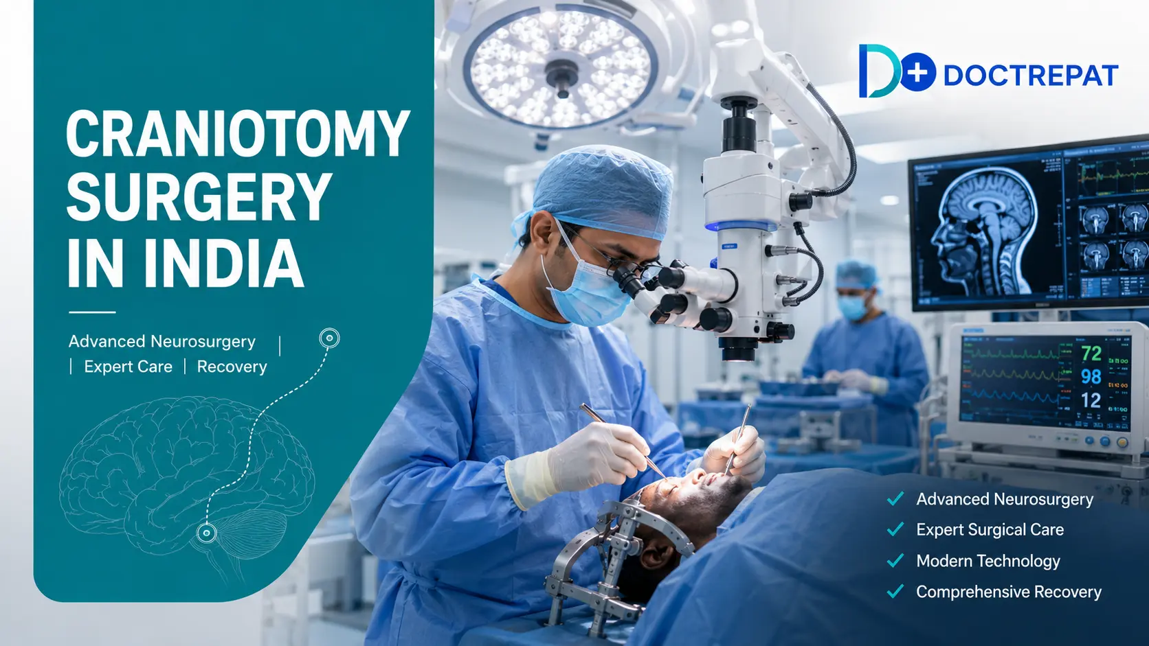
Craniotomy surgery in India for Ethiopian patients depends on the type of craniotomy, complexity, and underlying condition being treated. India offers advanced craniotomy procedures including awake craniotomy, minimally invasive keyhole craniotomy, and image-guided neurosurgery at NABH and JCI accredited hospitals, with success rates of 90 to 95% for planned craniotomies performed by internationally trained neurosurgeons.
Why This Guide Matters for Ethiopian Craniotomy Patients
Craniotomy is one of the most common major neurosurgical procedures performed globally, used to treat brain tumors, aneurysms, hemorrhages, epilepsy, and traumatic brain injuries. Ethiopian neurosurgical capacity remains limited, with Tikur Anbessa Specialized Hospital and Menelik II Referral Hospital in Addis Ababa performing basic craniotomies but lacking access to advanced neurosurgical technology.
Awake craniotomy, minimally invasive keyhole surgery, intraoperative MRI, neuronavigation systems, and specialized skull base surgery are largely unavailable in Ethiopia. Complex craniotomies for tumors near critical brain areas, aneurysms in difficult locations, or skull base pathologies typically require treatment abroad. India delivers world-class outcomes with over 15,000 craniotomies performed annually at leading neurosurgical centers.
Major Indian neurosurgical hospitals have decades of experience treating African patients, with Amharic interpreter services, dedicated international patient departments, and specialized neuro-rehabilitation programs.
What is a craniotomy?
A craniotomy is a surgical procedure where a section of the skull, called a bone flap, is temporarily removed to give the neurosurgeon access to the brain. After the underlying condition is treated, the bone flap is replaced and secured with small titanium plates and screws. This differs from a craniectomy, where the bone flap is not immediately replaced, usually because of brain swelling that requires additional room.
Craniotomy itself is not a treatment, it is the surgical access route that allows neurosurgeons to remove brain tumors, clip aneurysms, evacuate blood clots, or treat epilepsy. In Ethiopia, the Amharic terms commonly used are ክራንዮቶሚ (kraniyotomi) for craniotomy and የአንጎል ቀዶ ጥገና (ye'angol k'edo t'gena) for brain surgery in general.
Modern craniotomy is highly refined and precise. Depending on complexity, the procedure typically takes 3 to 8 hours and involves specialized microsurgical techniques using operating microscopes, neuronavigation systems, and intraoperative monitoring to protect critical brain functions.
What are the main types of craniotomy?
The main types of craniotomy include standard craniotomy (larger bone flap for major access), minimally invasive or keyhole craniotomy (smaller opening for precise access with less trauma), awake craniotomy (patient conscious during surgery to protect speech and motor functions), stereotactic craniotomy (image-guided precise targeting), and endoscopic craniotomy (camera-assisted, less invasive access).
Approach-based types include pterional craniotomy (the most common approach, used for aneurysms and frontal tumors), suboccipital craniotomy (accessing the back of the brain and cerebellum), and skull base craniotomy (specialized techniques for tumors at the base of the skull). The neurosurgeon selects the craniotomy type based on tumor location, patient anatomy, and specific goals of surgery.
What conditions require craniotomy?
Craniotomy is used to treat a wide range of brain conditions, most commonly brain tumors (meningioma, glioma, glioblastoma, pituitary adenoma, metastatic tumors), brain aneurysms (surgical clipping), and brain hemorrhages (intracerebral, subdural, or epidural bleeding). Other indications include arteriovenous malformations (AVMs), traumatic brain injury with significant bleeding, epilepsy surgery for focal seizure control, brain abscess drainage, Chiari malformation, and microvascular decompression for trigeminal neuralgia.
Ethiopian patients most commonly require craniotomy for brain tumors and aneurysms, both of which benefit significantly from the advanced neurosurgical technology and multidisciplinary expertise available at Indian centers. Time-sensitive conditions like ruptured aneurysms and acute hemorrhages require emergency evaluation, while planned procedures for tumors and epilepsy allow a 2 to 3 week window for optimal planning.
How is a craniotomy performed?
Craniotomy begins with detailed pre-surgical imaging including MRI with contrast, MR angiography for vascular conditions, and functional MRI when the tumor is near speech or motor areas. Neuronavigation software creates a 3D map of the patient's brain that guides the surgeon in real time during surgery.
The procedure involves general or awake anesthesia (depending on tumor location), a scalp incision that follows natural hair patterns to minimize visibility, creation of the bone flap using specialized surgical drills, opening of the dura (the brain's protective covering), and microsurgical treatment of the underlying condition. The operating microscope provides magnified visualization, ultrasonic aspirators (CUSA) allow precise tumor removal, and intraoperative neurophysiological monitoring tracks nerve function throughout the procedure. Once the surgical goal is achieved, the dura is closed, the bone flap is replaced and secured with titanium plates, and the scalp is closed with sutures or staples.
Total surgery time ranges from 3 to 8 hours depending on complexity. Patients spend 1 to 2 days in the ICU for neurological monitoring, followed by 5 to 8 days on the neurosurgery ward, for a total hospital stay of 5 to 10 days.
What is awake craniotomy?
Awake craniotomy is a specialized technique where the patient is conscious during critical portions of the surgery, allowing the neurosurgeon to test brain function in real time. The scalp is numbed with local anesthesia (the brain itself has no pain receptors), and the patient is sedated during opening and closing but woken for the microsurgical portion. The patient may be asked to speak, count, name objects, or move fingers while the surgeon operates near critical brain areas.
Awake craniotomy is used when the tumor is located near speech centers, motor areas, or memory regions, and for epilepsy surgery where preserving function is as important as removing abnormal tissue. The technique dramatically improves outcomes: patients experience fewer neurological deficits, greater tumor removal, and faster recovery compared to standard craniotomy for tumors in critical brain regions.
Awake craniotomy is available at all major Indian neurosurgical centers with dedicated teams including neurosurgeons, neuroanesthesiologists, neuropsychologists, and speech therapists. Success rates for awake craniotomy in India are 93 to 96%, matching leading Western centers.
What are the risks and complications of craniotomy?
All craniotomies carry surgical risks including bleeding, infection, anesthesia complications, and blood clots. Neurological risks specific to brain surgery include stroke, seizures, neurological deficits (weakness, speech difficulties, vision or memory changes), and cognitive changes. Additional risks may include cerebrospinal fluid (CSF) leak, hydrocephalus (fluid buildup in the brain), and swelling requiring additional intervention.
Risk levels depend heavily on the location and nature of the underlying condition, the patient's overall health, and the experience of the surgical team. Experienced neurosurgical centers with high case volumes consistently achieve lower complication rates. Modern techniques including neuronavigation, intraoperative monitoring, awake craniotomy for critical regions, and minimally invasive approaches have significantly reduced risks over the past two decades. At leading Indian centers, complication-free recovery is achieved in 85 to 92% of cases.
Which Indian hospitals are best for craniotomy for Ethiopian patients?
Leading Indian hospitals for craniotomy surgery for Ethiopian patients include BLK Max Super Speciality Hospital Delhi, Fortis Memorial Research Institute Gurugram, Artemis Hospital, Manipal Hospital, and Paras Hospital. All operate comprehensive neurosurgery programs with advanced technology including intraoperative MRI, neuronavigation systems, and dedicated neuro-ICUs.
BLK Max Super Speciality Hospital operates one of Delhi's most established neurosurgery units with a dedicated awake craniotomy program and complex skull base surgery capability. Fortis Memorial Research Institute houses advanced neurosurgical technology including intraoperative MRI, allowing real-time imaging during surgery to ensure complete tumor removal. Artemis Hospital operates a comprehensive Neurosciences Institute with subspecialty expertise in vascular neurosurgery, epilepsy surgery, and pediatric neurosurgery. Manipal Hospital runs high-volume neurosurgery programs with extensive experience treating African patients. Paras Hospital provides advanced neurosurgical care with specialized skull base and functional neurosurgery programs.
Hospital selection depends on the specific condition, procedure complexity, need for awake craniotomy or specialized technology, and family logistics. DocTrePat coordinates hospital matching through case review with the treating neurosurgical team.
What is the success rate of craniotomy in India?
Success rates for craniotomy in India match leading Western centers, with 90 to 95% success rates for planned craniotomies performed by experienced neurosurgeons. Outcomes depend significantly on the underlying condition, tumor location, and complexity of the procedure.
Success rates are highest for benign tumors, aneurysm clipping, and awake craniotomy for tumors in critical brain regions. Emergency craniotomy outcomes depend heavily on time to surgery and pre-operative neurological status. Indian outcomes match Western centers because the same neurosurgical techniques, technology, and evidence-based protocols are used at accredited hospitals.
What is recovery like after a craniotomy?
Immediate recovery after craniotomy involves 24 to 48 hours in the neuro-ICU for close neurological monitoring, pain management, and prevention of complications. Patients are typically alert and communicating within a few hours of surgery, though drowsiness and mild confusion are common initially. Early mobilization begins within 24 hours when possible.
The first month after surgery focuses on wound healing, gradual return to normal activities, and management of any temporary neurological symptoms. Most patients experience some fatigue, mild headaches, and occasional dizziness during this period. Sutures or staples are removed 7 to 10 days after surgery, and the surgical wound typically heals well with minimal visible scarring since incisions follow hair patterns.
By 3 to 6 months post-surgery, most patients have returned to normal daily activities including work or school. Physical therapy, occupational therapy, and speech therapy are provided when needed to address any temporary functional deficits. Cognitive rehabilitation may be recommended for patients who had surgery near memory or executive function areas.
Long-term follow-up involves periodic MRI or CT imaging to monitor for tumor recurrence (in cancer cases), aneurysm re-formation, or other complications. Many follow-up visits can be conducted through teleconsultation with the Indian neurosurgical team while the patient is back in Ethiopia, with periodic return visits for imaging.
How does an Ethiopian patient travel to India for craniotomy?
First, DocTrePat receives medical reports including MRI, CT scans, angiography (if applicable), and clinical notes, and routes them to an Indian neurosurgeon for review. A surgical plan and hospital invitation letter are provided within 48 hours. For emergency cases including ruptured aneurysms and acute hemorrhages, expedited 24-hour case review is available with urgent hospital admission arrangements.
Second, the patient applies for an Indian medical visa at the Indian Embassy in Addis Ababa. Standard processing takes 5 to 10 working days, with expedited 2 to 3 day processing available for medical urgency. A companion visa for one attendant is essential because post-operative assistance is required. Multi-entry visas are recommended for patients who may need follow-up visits.
Third, the patient flies Ethiopian Airlines from Addis Ababa Bole International Airport to Delhi, Mumbai, or Chennai depending on the chosen hospital. DocTrePat coordinates airport pickup with wheelchair or stretcher arrangements when needed, and direct transfer to the hospital.
Pre-operative testing and consultation typically require 2 to 3 days after arrival, with surgery scheduled within 3 to 7 days. Post-operative hospital stay is 5 to 10 days, followed by outpatient monitoring for 1 to 2 weeks before returning to Ethiopia. Total time in India: 3 to 4 weeks minimum for elective craniotomy, 4 to 6 weeks for complex procedures.
Get a Personalized Craniotomy Treatment Plan in India
Whether you need brain tumor surgery, aneurysm treatment, or another neurosurgical procedure, get a specialist second opinion and personalized treatment plan from experienced Indian neurosurgeons.
📱 WhatsApp: +91 96259 33363
Share Your Medical Reports & Get Expert Guidance →
Frequently Asked Questions
India offers several advantages over what is currently available in Ethiopia: awake craniotomy for tumors in critical brain areas, minimally invasive keyhole approaches, intraoperative MRI for real-time surgical guidance, neuronavigation systems, dedicated skull base surgery teams, comprehensive neuro-rehabilitation, and 24-hour multidisciplinary neuro-ICU care. Ethiopian neurosurgery at Tikur Anbessa and Menelik II provides basic craniotomy capability, but advanced technology and subspecialty expertise remain limited.
Most patients can safely return to Ethiopia 2 to 3 weeks after craniotomy surgery, after wound healing is confirmed and neurological status is stable. Complex cases or patients with post-operative complications may require longer stays of 4 to 6 weeks. Follow-up visits at 3 and 6 months can be planned in advance or replaced with teleconsultation and local imaging in Ethiopia.
Only a small area of hair around the incision site is typically shaved before surgery, and this hair grows back within 2 to 3 months. Modern neurosurgical technique minimizes hair removal and uses incisions that follow natural hair patterns to hide the scar. Full hair shaving is rarely required for standard craniotomy.
Yes, awake craniotomy is available at all major Indian neurosurgical centers including BLK Max, Fortis, Artemis, Manipal, and Paras hospitals. Dedicated awake craniotomy teams including neurosurgeons, neuroanesthesiologists, and neuropsychologists ensure safe execution. Success rates for awake craniotomy in India are 93 to 96%, matching leading Western centers.
Initial recovery in the hospital takes 5 to 10 days, followed by 3 to 6 weeks of continued healing at home. Most patients return to normal daily activities within 6 to 8 weeks. Complete recovery including full return to work, exercise, and driving typically takes 3 to 6 months depending on the underlying condition and any neurological deficits requiring rehabilitation.
Craniotomy surgery typically takes 3 to 8 hours depending on complexity. Simple procedures like biopsies or small tumor removals may take 3 to 4 hours, while complex skull base surgeries or AVM resections can take 6 to 8 hours or longer. Awake craniotomy typically takes 4 to 6 hours including patient wake-up periods for functional testing.
Yes, craniotomy surgery in India is safe when performed at accredited hospitals by experienced neurosurgeons. Success rates for planned craniotomies are 90 to 95%, and complication-free recovery is achieved in 85 to 92% of cases. Indian centers use the same neurosurgical techniques, technology, and evidence-based protocols as leading Western hospitals, and many neurosurgeons have international training and fellowship experience.
Our team is here to help you with personalized answers, treatment guidance, and free cost estimates. Reach out anytime, we’ll respond within 24 hours.
Get in Touch Now!
.webp)